How to Cite
Copyright notice

This work is licensed under a Creative Commons Attribution-NonCommercial 4.0 International License.
Copyright (c) 2021 Tobin Driscoll, Richard J. Braun, Carolyn G. Begley
Keywords
Abstract
Purpose: Fluorescence imaging is a valuable tool for studying tear film dynamics andcorneal staining. Automating the quantification of fluorescence images is a challenging necessary step for making connections to mathematical models. A significant partof the challenge is identifying the region of interest, specifically the cornea, for collected data with widely varying characteristics.
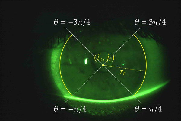
Methods: The gradient of pixel intensity at the cornea–sclera limbus is used as the objective of standard optimization to find a circle that best represents the cornea. Results of the optimization in one image are used as initial conditions in the next imageof a sequence. Additional initial conditions are chosen heuristically. The algorithm iscoded in open-source software.
Results: The algorithm was first applied to 514 videos of 26 normal subjects, for a total of over 87,000 images. Only in 12 of the videos does the standard deviation in thedetected corneal radius exceed 1% of the image height, and only 3 exceeded 2%. The algorithm was applied to a sample of images from a second study with 142 dry-eye subjects. Significant staining was present in a substantial number of these images. Visual inspection and statistical analysis show good resuls for both normal and dry-eye images.
Conclusion: The new algorithm is highly effective over a wide range of tear film andcorneal staining images collected at different times and locations.
References
Craig JP, Nichols KK, Akpek EK, Caery B, Dua HS, Joo CK, et al. TFOS DEWS II Definition and Classification Report. en. The Ocular Surface. TFOS International Dry Eye WorkShop (DEWS II), July 2017;15(3): 276–283. issn: 1542-0124. doi: 10/gbstrp.
Norn MS. Desiccation of the Precorneal Film. en. Acta Ophthalmologica, 1969;47(4): 881–889. issn: 1755-3768. doi: 10/dj95jq.
Cho P, BrownB, Chan I,Conway R, Yap M. Reliability of the Tear Break-Up Time Technique of Assessing Tear Stability and the Locations of the Tear Break-Up in Hong Kong Chinese: en.Optometry and Vision Science, Nov. 1992;69(11): 879–885. issn: 1040-5488. doi: 10/fb4kjv.
Lemp MA. Report of the National Eye Institute/Industry Workshop on Clinical Trials in Dry Eyes. eng. The CLAO journal: official publication of the Contact Lens Association of Ophthalmologists, Inc, Oct. 1995;21(4): 221–232. issn: 0733-8902.
Bron AJ, Evans VE, Smith JA. Grading of Corneal and Conjunctival Staining in the Context of Other Dry Eye Tests. eng. Cornea, Oct. 2003;22(7): 640–650. issn: 0277-3740. doi: 10/d648xt.
Whitcher JP, Shiboski CH, Shiboski SC, Heidenreich AM, Kitagawa K, Zhang S, et al. A Simplified Quantitative Method for Assessing Keratoconjunctivitis Sicca from the Sjögren’s Syndrome International Registry. eng. American Journal of Ophthalmology, Mar. 2010;149(3): 405–415. issn: 1879-1891. doi:10/ckdjf2.
Terry RL, Schnider CM, Holden BA, Cornish R, Grant T, Sweeney D, et al. CCLRU Standards for Success of Daily and Extended Wear Contact Lenses. eng. Optometry and Vision Science: Official Publication of the American Academy of Optometry, Mar. 1993;70(3): 234–243. issn: 1040-5488. doi: 10/dw2dwf.
Norn MS. Tear Film Breakup Time: A Review. The Preocular Tear Film in Health, Disease and Contact Lens Wear. Lubbock, TX: Dye Eye Institute, 1986; 52–56.
Liu H, Begley CG, Chalmers R, Wilson G, Srinivas SP, Wilkinson JA. Temporal Progression and Spatial Repeatability of Tear Breakup. en-US. Optometry and Vision Science, Oct. 2006;83(10): 723–730. issn: 1538-9235. doi: 10/bsp8tf.
Amparo F, Wang H, Yin J, Marmalidou A, Dana R. Evaluating Corneal Fluorescein Staining Using a Novel Automated Method. eng. Investigative Ophthalmology & Visual Science, May 2017;58(6):BIO168–BIO173. issn: 1552-5783. doi: 10/gbmdx3.
Pritchard N, Young G, Coleman S, Hunt C. Subjective and Objective Measures of Corneal Staining Related to Multipurpose Care Systems. eng. Contact Lens & Anterior Eye: The Journal of the British Contact Lens Association, Mar. 2003;26(1): 3–9. issn: 1367-0484. doi: 10/bq32wm.
Wolffsohn JS, Purslow C. Clinical Monitoring of Ocular Physiology Using Digital Image Analysis. en. Contact Lens and Anterior Eye, Mar. 2003;26(1): 27–35. issn: 13670484. doi: 10/c26rht.
Tan B, Zhou Y, Svitova T, Lin MC. Objective Quantification of Fluorescence Intensity on the Corneal Surface Using a Modified Slit-Lamp Technique. eng. Eye & Contact Lens, May 2013;39(3): 239–246. issn: 1542-233X. doi: 10/f4xc3x.
Chun YS, Yoon WB, Kim KG, Park IK. Objective Assessment of Corneal Staining Using Digital Image Analysis. en. Investigative Ophthalmology & Visual Science, Dec. 2014;55(12): 7896–7903. issn: 1552-5783. doi: 10/f6txkr.
Pellegrini M, Bernabei F, Moscardelli F, Vagge A, Scotto R, Bovone C, et al. Assessment of Corneal Fluorescein Staining in Different Dry Eye Subtypes Using Digital Image Analysis. eng. Translational Vision Science & Technology, Nov. 2019;8(6): 34. issn: 2164-2591. doi: 10/ghwn39.
Sher I, Tzameret A, Szalapak AM, Carmeli T, Derazne E, Avni-Zauberman N, et al. Multimodal Assessment of Corneal Erosions Using Optical Coherence Tomography and Automated Grading of Fluorescein Staining in a Rabbit Dry Eye Model. eng. Translational Vision Science & Technology, Jan. 2019;8(1): 27. issn: 2164-2591. doi: 10/ghwn38.
Vyas AH, Mehta MA. A Comprehensive Survey on Image Modality Based Computerized Dry Eye Disease Detection Techniques. Advances in Science, Technology and Engineering Systems Journal, 2020;5(2): 748–756. issn: 24156698, 24156698. doi: 10/ghwn37.
Ramos L, Barreira N, Mosquera A, Penedo MG, Yebra-Pimentel E, García-Resúa C. Analysis of Parameters for the Automatic Computation of the Tear Film Break-up Time Test Based on CCLRU Standards. en. Computer Methods and Programs in Biomedicine, Mar. 2014;113(3): 715–724. issn: 0169-2607. doi: 10/f5tgrc.
RamosL, Barreira N,Pena-Verdeal H, Giráldez MJ, Yebra-Pimentel E.Computational Approach for Tear Film Assessment Based on Break-up Dynamics. en. Biosystems Engineering. Innovations in Medicine and Healthcare, Oct. 2015;138, 90–103. issn: 1537-5110. doi: 10/f7stvd.
Remeseiro B, Mosquera A,Penedo MG, Garcia-Resúa C. Tear Film Maps Based on the Lipid Interference Patterns: Proceedings of the 6th International Conference on Agents and Artificial Intelligence. ESEO, Angers, Loire Valley, France: SCITEPRESS - Science and and Technology Publications, 2014; 732–739. isbn: 978-989-758-015-4 978-989-758-016-1. doi: 10/ghwn4c.
Daugman J. How Iris Recognition Works. IEEE Transactions on Circuits and Systems for Video Technology, Jan. 2004;14(1): 21–30. issn: 1558-2205. doi: 10/bwxqsn.
Su TY, Liu ZY, Chen DY. Tear Film Break-Up Time Measurement Using Deep Convolutional Neural Networks for Screening Dry Eye Disease. IEEE Sensors Journal, Aug. 2018;18(16): 6857–6862. issn: 2379-9153. doi: 10.1109/jsen.2018.2850940.
Driscoll TA. Automatic cornea detection in fluorescence images (0.1.0). 2021; doi: 10.5281/zenodo.5495707.
Awisi-Gyau D, Begley CG, Situ P, Simpson TL. Changes in Corneal Detection Thresholds After Repeated Tear Film Instability. en. Investigative Opthalmology & Visual Science, Oct. 2019;60(13): 4234. issn: 1552-5783. doi: 10/ghwn4b.
Simpson T, Begley CG, Situ P, Feng, Nelson JD, Caery B, et al. Canonical Grading Scales of Corneal and Conjunctival Staining Based on Psychophysical and Physical Attributes. Translational Vision Science and Technology,in review.
Duke-Elder S, Wybar KC. The Cornea. The Anatomy of the Visual System. 2. Systemof Ophthalmology. London: Henry Kimpton, 1961; 92–94.
Khng C, Osher RH. Evaluation of the Relationship between Corneal Diameter and Lens Diameter. en-US. Journal of Cataract & Refractive Surgery, Mar. 2008;34(3): 475–479. issn: 0886-3350. doi: 10/dvknbj.