How to Cite
Copyright notice

This work is licensed under a Creative Commons Attribution-NonCommercial 4.0 International License.
Copyright (c) 2021 Gabor Hollo
Keywords
Abstract
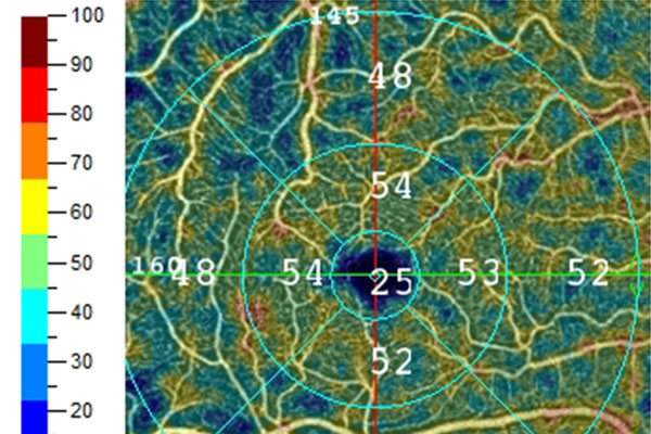
Background: In ophthalmology, thickness and vessel density (VD) measurements for the 6 x 6 mm inner macular retinal area have received increasing attention in glaucomatous progression research. For this area, the Angiovue optical coherence tomography system introduced a 304 x 304 A/B scans function (classic Angio Retina scan) in 2014, and a 400 x 400 A/B scans function (high-definition [HD] Angio Retina scan) in 2017. These scan types cannot be used in combination for the software provided for progression analysis.
Purpose: Since losing data for 3 years may negatively influence progression analysis, we investigated whether clinically significant differences exist between consecutive measurements acquired with these scan types on the same eyes.
Methods: As a part of our noninterventional prospective glaucoma imaging study, primary-open-angle glaucoma patients (POAG group), and ocular hypertensive and healthy control participants (structurally undamaged group) were imaged
using both the classic and the HD Angio Retina scans, respectively, without changing the patients’ position. High-quality images were obtained on 12 POAG eyes of 12 consecutive POAG patients, and 10 healthy and ocular hypertensive eyes of 10 consecutive participants before the data collection had to be suspended due to the new coronavirus epidemic.
Results: For Early Treatment Diabetic Retinopathy Study image area, the mean difference (classic minus HD value) was 0.02 ± 0.37 μm for inner retinal thickness (P = 0.869) and 0.33 ± 1.33 % (P = 0.452) for superficial capillary VD in the structurally normal group (between-methods difference: ≤ 0.8% of the respective normal value). In the POAG group, the corresponding figures were -0.07 ± 1.22 μm for inner retinal thickness (P = 0.854; between-methods difference: 0.6% of the normal value) and 1.12 ± 2.58 % for superficial capillary VD (P = 0.158; classic scan value minus HD scan value: 1.12 ± 2.58 %; 2.3% of the normal value).
Conclusion: Our results suggest that combined use of thickness and VD values for structurally normal eyes and thickness values for POAG eyes derived from classic and HD scans, respectively, for progression analysis can be reasonable since the differences between the corresponding values are small. However, combining the corresponding VD parameters for POAG eyes is useful only when the follow-up time before the scan type change is long enough to counterbalance the effect of the change on the result.
References
Tham Y-C, Li X, Wong TY, et al. Global prevalence of glaucoma and projections of glaucoma burden through 2040: a systematic review and meta-analysis. Ophthalmology 2014;121:2081-90. https://doi.org/10.1016/j.ophtha.2014.05.013.
Pinto AL, Willekens K, Van Keer K, et al. Ocular blood flow in glaucoma - the Leuven Eye Study. Acta Ophthalmol. 2016;94:592-598. https://doi.org/10.1111/aos.12962.
Flammer J, Orgül S, Costa VP, et al. The impact of ocular blood flow in glaucoma. Prog Retin Eye Res. 2002;21:359-93. https://doi.org/10.1016/s1350-9462(02)00008-3.
Hou H, Moghimi S, Zangwill LM, et al. Macula vessel density and thickness in early primary open-angle glaucoma. Am J Ophthalmol. 2019;199:120-132. https://doi.org/10.1016/j.ajo.2018.11.012
Moghimi S, Zangwill LM, Penteado RC, et al. Macular and optic nerve head vessel density and progressive retinal nerve fiber layer loss in glaucoma. Ophthalmology. 2018;125:1720-1728. https://doi.org/10.1016/j.ophtha.2018.05.006
Hou H, Moghimi S, Proudfoot JA, et al. Ganglion cell complex thickness and macular vessel density loss in primary open-angle glaucoma. Ophthalmology. 2020;127:1043-1052. https://doi.org/10.1016/j.ophtha.2019.12.030
Holló G. Comparison of thickness – function and vessel density – function relationship in the superior and inferior macula, and the superotemporal and inferotemporal peripapillary sectors. J Glaucoma. 2020;29:168-174. https://doi.org/10.1097/IJG.0000000000001441
Optovue Inc. RTVue XR Avanti System. User Manual International Software Version 2017.1. Release date: 11/2017.
Hodapp E, Parrish RK, Anderson DR. Clinical Decisions in Glaucoma. St Louis, MO: Mosby; 1993. pp. 52–61.
Ye C, Wang X, Yu MC, et al. Progression of macular vessel density in primary open-angle glaucoma: a longitudinal study. Am J Ophthalmol. 2020;223:259-266. https://doi.org/10.1016/j.ajo.2020.10.008